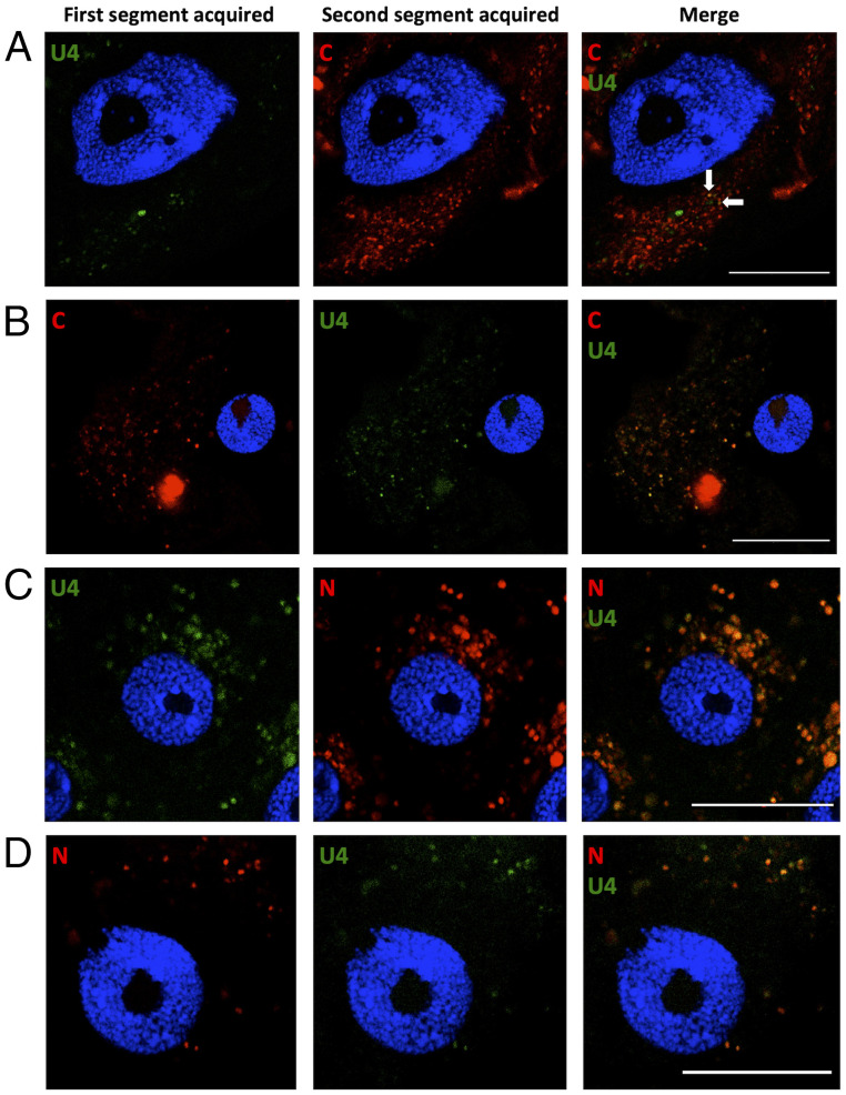
Fig. 4.
Localization of sequentially acquired DNA segments of FBNSV in aphid AMG cells. Viral DNA is labeled by FISH in AMG cells of viruliferous aphids and observed by confocal microscopy. The green probe targets segment U4, and the red probes target either C or N in the corresponding panels. The accumulation of FBNSV DNA was similarly revealed in all observed cells (>10 cells per midgut) from 16, 10, 6, and 14 viruliferous aphids for A, B, C, and D, respectively. A representative image of each case is shown to illustrate the results. A and B represent the sequential acquisition of segments C/U4, and C and D represent that of segments N/U4, in the indicated sequential order. The white arrows show two foci containing both C and U4 segments. All images correspond to single optical sections. Cell nuclei are stained with DAPI (blue). Scale bar, 25 µm.

