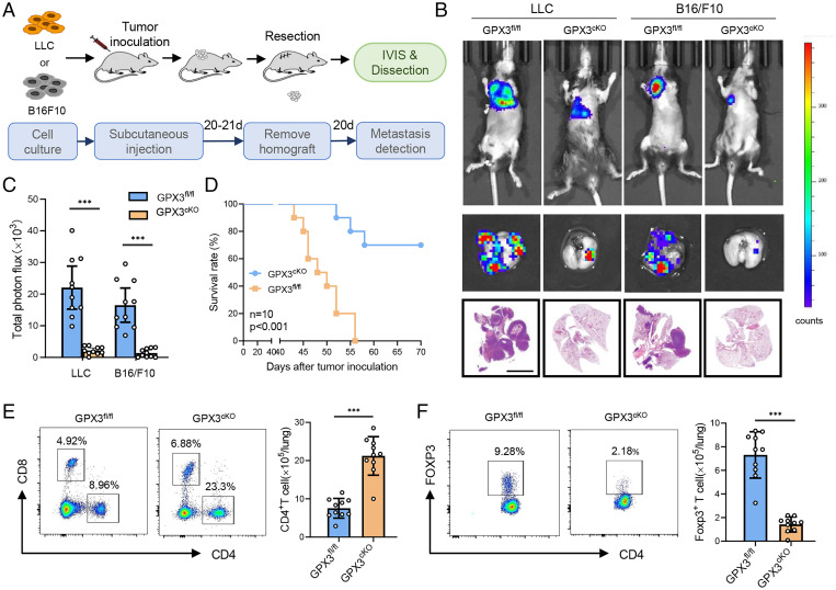
Fig. 2.
CKO of GPX3 in AT2 cells inhibits tumor metastasis. (A) Diagram for the mouse model of spontaneous lung metastasis of LLC and B16/F10. IVIS, in vivo imaging system. (B and C) Representative images (B) and quantitative analysis (C) of lung metastasis of GPX3fl/fl or GPX3cKO mice detected by luciferase-based bioluminescence imaging or by H&E-stained lung sections and quantification of lung metastatic foci of GPX3fl/fl or GPX3cKO mice (n = 10) 40 d after LLC or B16/F10 inoculation. Scale bar, 5 mm. (D) Survival of GPX3fl/fl or GPX3cKO mice (n = 10 each) after LLC inoculation. Kaplan-Meier test. (E and F) Flow cytometry analysis of the proportions and absolute numbers of CD4+ T cells, CD8+ T cells (E), and CD4+ Foxp3+ Treg cells (F) in the lungs of GPX3fl/fl or GPX3cKO mice 14 d after LLC inoculation. Data are mean ± SD of one representative experiment. Similar results were seen in three independent experiments. Unpaired Student’s t tests unless noted. ***P < 0.001.

