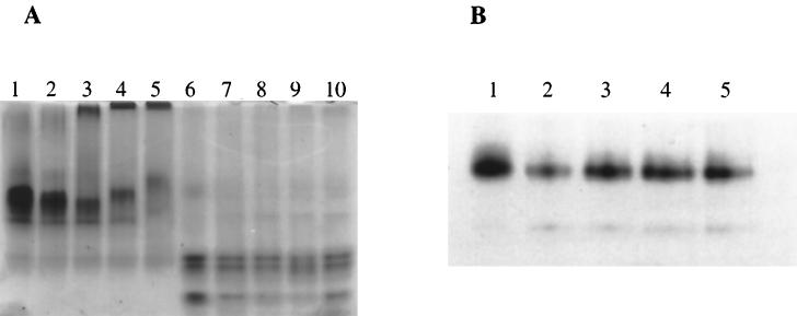
FIG. 7.
Reconstitution of proteins in lipid vesicles. Proteins were reconstituted into lipid vesicles as described in Materials and Methods. The vesicles were analyzed by native 8% PAGE. (A) PBP 2a* and the TP domain. Proteins (10 μg) were stained with Coomassie blue. Lanes: 1, PBP 2a* in 3% CHAPS; 2 to 5, PBP 2a* in the presence of molar protein/lipid ratios of 0, 1/50, 1/200, and 1/500, respectively; 6, TP in 3% CHAPS; 7 to 10, TP in the presence of molar protein/lipid ratios of 0, 1/50, 1/200, and 1/500, respectively. (B) PBP 2a* thrombin-digested product. Protein detection (0.7 μg) was performed by [3H]benzylpenicillin labelling. Lanes: 1, thrombin-digested PBP 2a* in 3% CHAPS; 2 to 5, thrombin-digested PBP 2a* in the presence of molar protein/lipid ratios of 0, 1/200, 1/500, and 1/1,000, respectively.

