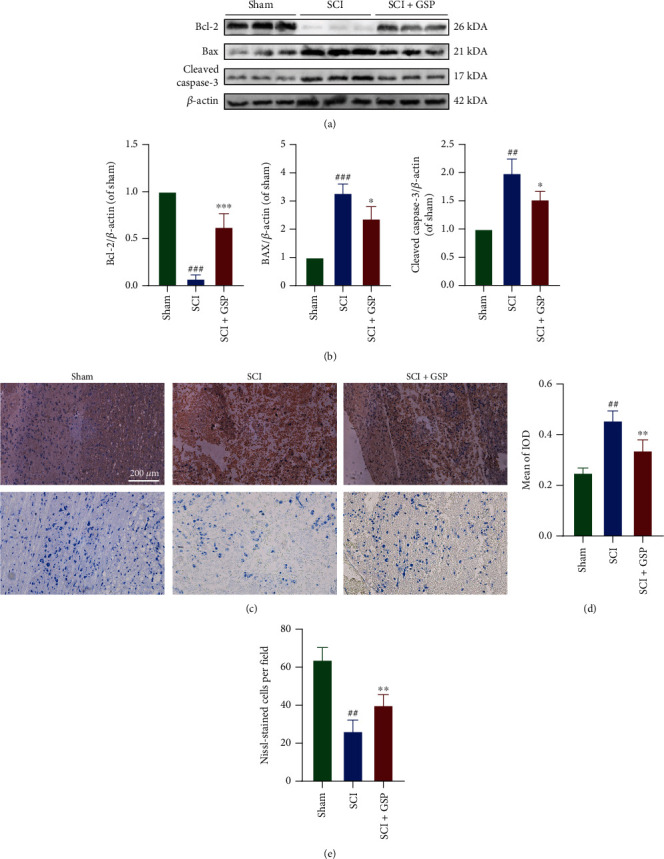
Figure 5.

GSP inhibit apoptosis in vivo. (a, b) Representative WB and quantitative data of Bax, Bcl-2, and cleaved caspase-3 in the different groups at 14 dpi (n = 3 rats in each group). (c, d) Representative IHC staining and Nissl staining in the different groups at 14 dpi. (d) Quantitative data of cleaved caspase-3. (n = 3, with 5 images for each rat). (e) Quantitative data of the number of Nissl-stained cells at 14 days after SCI (n = 3 rats in each group). ##p < 0.01 or ###p < 0.001 vs. sham group. ∗p < 0.05, ∗∗p < 0.01, or ∗∗∗p < 0.001 vs. SCI group.
