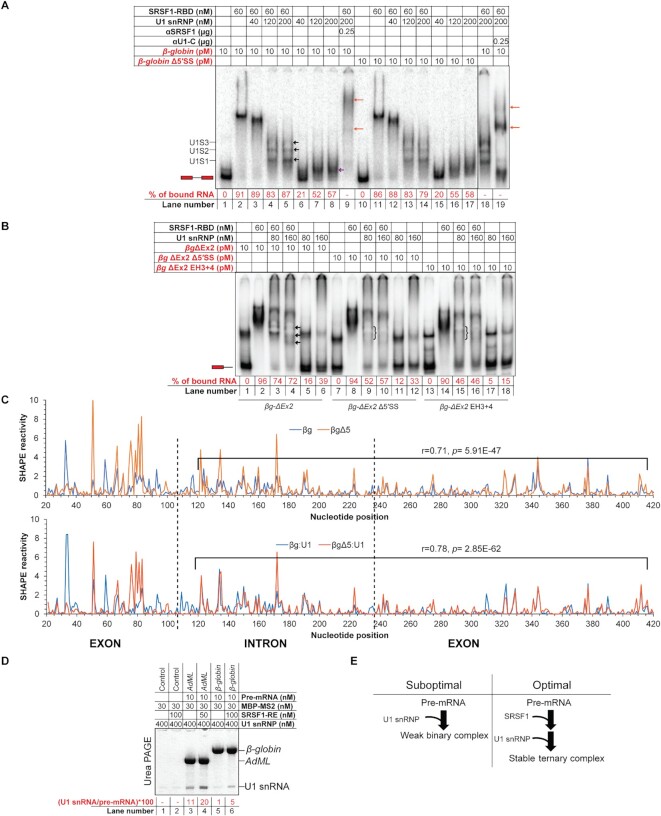
Figure 1.
U1 snRNP recognizes the pre-mRNA 3D structural scaffold in β-globin. (A) β-globin forms ternary complexes with SRSF1-RBD and U1 snRNP, which migrate primarily as three major bands (marked with black arrows in lane 5 and labeled as U1S1, U1S2 and U1S3 on the side of the gel); U1 snRNP also binds free β-globin (marked with a violet arrow in lane 8); the ternary complex is super-shifted with αSRSF1 (lane 9, marked with orange arrows) and αU1-C (lanes 18 and 19); slightly more smeary complexes of similar migration patterns were formed with β-globin Δ5′SS (lanes 10–17); red script indicates radiolabeled components; the position of the free probe is indicated with an exon-intron-exon schematic; percentage of upshifted β-globin probe is indicated below each lane. (B) βg-ΔEx2 (β-globin lacking the 3′ exon) formed U1 snRNP-dependent complexes in the presence of 60 nM SRSF1-RBD (the complexes are marked with arrows in lane 4); these U1 snRNP-dependent complexes were significantly weakened for βg-ΔEx2 with 5′SS mutations (βg-ΔEx2 Δ5′SS) and βg-ΔEx2 with hybridization-mutation immediately upstream of the 5′SS (βg-ΔEx2 EH3 + 4) (the position of the weakened complexes are marked with curved brackets in lanes 9 and 15); U1 snRNP shows no well-defined interaction with the free RNAs (lanes 5, 6, 11, 12, 17, 18); the position of the free probe is indicated with an exon-intron schematic. (C) Overlaid SHAPE reactivity of protein-free β-globin WT and its 5′SS mutant (Δ5) (top) and U1 snRNP-bound β-globin WT and U1 snRNP-bound β-globin 5′SS-mutant (bottom); the segments showing a moderate correlation of SHAPE reactivity are marked in each plot and the corresponding r and p values are indicated; exon-intron boundaries are demarcated with dotted vertical lines. (D) Amylose pull-down assay showing enhancement of co-purification of U1 snRNP (indicated by corresponding U1 snRNA level) with AdML and β-globin in the presence of SRSF1-RE by urea PAGE. (E) Summary flow chart: U1 snRNP specifically recognizes a global 3D structural scaffold of β-globin and SRSF1 enhances this interaction.

