Figure 1.
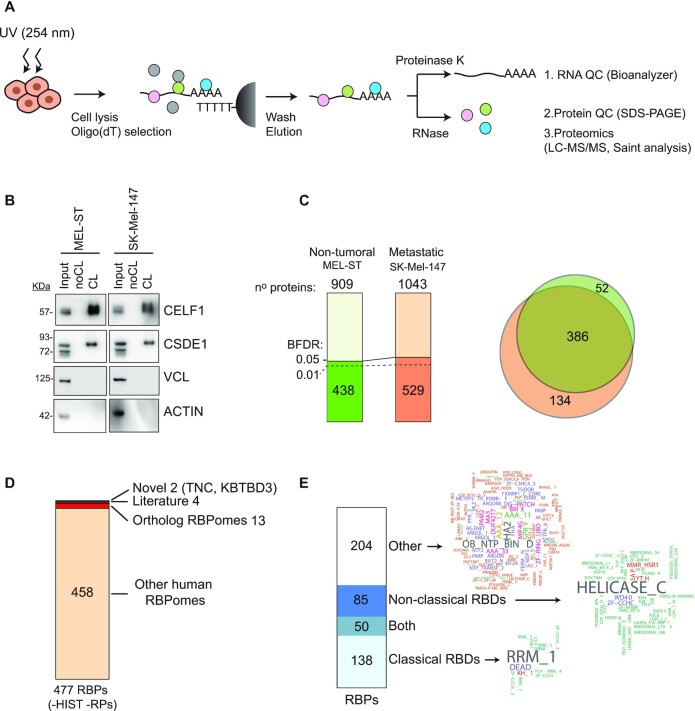
Characterization of the melanoma RBPome. (A) Schematic representation of the RNA interactome capture (RIC) procedure. (B) RBP enrichment assessed by western blot. (C) Number of total and significant (BFDR ≤ 0.05) proteins recovered in RIC eluates. Most significant proteins were also recovered with a BFDR ≤0.01 (dashed line). Significance was calculated using SAINTexpress by comparing crosslinked versus non-crosslinked conditions. The overlap of significant RBPs in both cell lines is shown on the right. (D) Comparison of the melanoma RBPome (excluding histones and ribosomal proteins) with previously published RBPomes. (E) Analysis of RNA binding domains (RBDs) present in the melanoma RBPome. Domain word clouds indicate the frequency of protein domains in the data sets.

