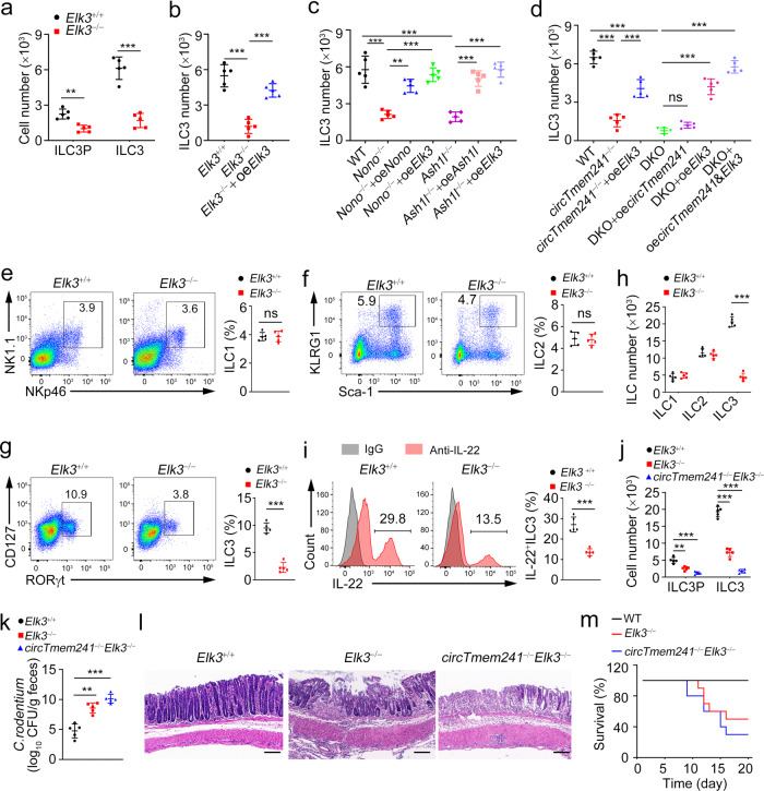
Fig. 7. Elk3 knockout impairs ILC3 commitment and anti-bacterial immunity.
a–d Indicated ILCPs were isolated and conducted in vitro differentiation assay with OP9-DL1 feeder cells. ILC3Ps were analyzed 7 days later and ILC3s were analyzed 14 days later by FACS. n = 5 for each group. e–g ILC1s (CD3−CD19−CD45+CD127+ gated), ILC2s (CD3−CD19−CD127+CD90+ gated) and ILC3s (CD3−CD19−CD45lo gated) were analyzed in small intestines of Elk3+/+ and Elk3−/− mice by FACS. n = 5 for each group. h Numbers of indicated ILCs in e–g were calculated. n = 5 for each group. i LPLs from Elk3+/+ and Elk3−/− mouse intestines were stimulated by IL-23 for 4 h, followed by IL-22+ ILC3 detection with FACS. j ILC3Ps and ILC3s from Elk3+/+, Elk3−/−, and circTmem241−/−Elk3−/− mice were analyzed by FACS, and indicated cell numbers were calculated. n = 5 for each group. k CFUs in feces of Elk3+/+, Elk3−/− and circTmem241−/−Elk3−/− mice were measured after C. rodentium infection for 6 days. n = 5 for each group. l Colon tissues from Elk3+/+, Elk3−/− and circTmem241−/−Elk3−/− mice were analyzed by H&E staining. Scale bars, 100 μm. m Survival rates of indicated mice were measured after infection with C. rodentium. n = 10 for each group. **P < 0.01 and ***P < 0.001. Data were analyzed by an unpaired two-side Student’s t test and shown as means ± SD. Data are representative of at least three independent experiments. Source data are provided as a Source Data file.

