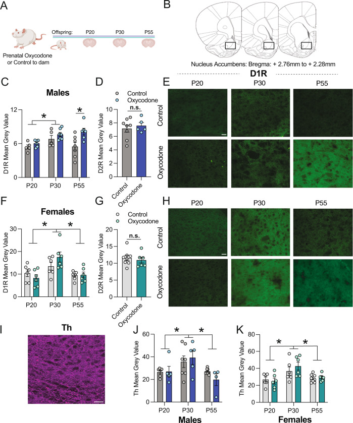
Fig. 2. Prenatal oxycodone exposure increases D1R density in the NAc in adulthood in males but not females.
A Schematic of timeline for treatment and tissue collection. B Rat brain atlas images delineating NAc region where D1R, D2R, and Th were quantified (based on Paxinos and Watson Rat Brain Atlas). C In males, D1R mean grey value differed significantly with age, and was higher at P55 in oxycodone-exposed males as compared to control. D D2R did not differ with treatment at P55 in males. E Representative 20x images of D1R staining in the NAc of males, scale bar = 27 microns. F D1R peaked at P30 in both control and oxycodone-exposed females, but did not differ with treatment. G D2R did not differ with treatment at P55 in females. H Representative 20x images of D1R staining in the NAc of females, scale bar = 27 microns. I Representative 20x image of Th staining in the NAc of males, scale bar = 27 microns. J Th peaked at P30 in both control and oxycodone exposed males and females (K), but did not differ with treatment. Data represent Mean +/− SEM, *p = 0.05.

