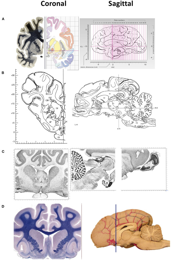
Figure 1.
Representative images of two-dimensional sections of the brain, coronal (right) and sagittal (left) sectons of brains of various species. (A) Human brain. Coronal sections show the fiber tracts in the left panel and schematic diagrams in the right panel. The sagittal section shows the plane of the 69 sections depicted in the atlas. The intercommisural line (ICL) and the vertical line (VCA) pass through the center of the anterior and posterior commissure, and the center of the anterior commissure, respectively. (B) Pig brain. Coronal and sagittal sections in this example show the coordinates 14.50 mm ahead of the posterior commissure and 4.00 mm laterally from the midsagittal plane, respectively. (C) Cat brain. Coronal and sagittal sections are examples of Nissl stained sections from the adult cat (Felis Catus). (D) Dog brain. Coronal and sagittal sections are shown in the maps of the whole brain from a 5 month old dog (Canis Lupus). The blue line shows the levels at the frontal lobe Images in (A) are reproduced from the human brain website https://www.thehumanbrain.info/brain/sections.php. Retrieved May 12, 2022. Images in (B) are reproduced from the Stereotaxic atlas of the pig brain by Felix et al. (1) with permission from Elsevier. Images (C,D) are reproduced from the brain maps website http://brainmaps.org/ajax-viewer.phpdatid=32&sname=p099-100, retrieved on May 13, 2022.

