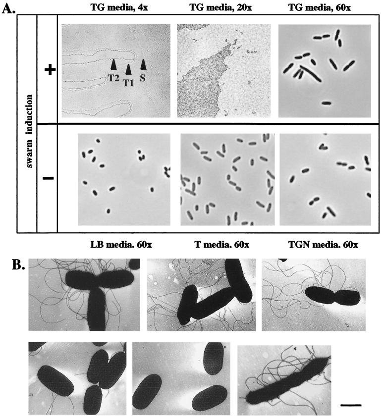
FIG. 3.
Images of colony and cellular morphology of swarming bacteria suggest that Y. enterocolitica differentiates into cells that cooperatively migrate over agar surfaces. (A) The top row shows images of the morphology of an advancing colony of bacteria grown on a TG plate at 26°C, conditions that induce swarming motility. (Top left) Image of a colony at ×4 magnification; S indicates the location of the slime layer preceding the front of migrating colony; T1 indicates the first terrace of migrating cells in the swarming colony; T2 indicates the location of the second terrace of cells that appear to overlay cells forming the first terrace. (Top middle) Image of the same colony at ×20 magnification, showing rafts of cells that often advance ahead of a colony. (Top right) Image of elongated cells resuspended from a swarming colony taken at ×60 magnification. The bottom row shows images (at ×60 magnification) of bacteria grown on LB plates, T plates, or T plates containing 100 mM glucose and 1% NaCl (TGN) at 26°C, conditions that influence swimming motility but do not induce swarming motility. (B) Electron micrographs of bacteria isolated under conditions that influence motility. Bacteria were isolated from agar plates grown for 16 h at 26°C. (Top left) Cells isolated under conditions that induce swimming motility on T medium. (Top middle) Cells isolated under swarm-induced conditions on TG medium. (Top right) A single swarm cell that appears to be dedifferentiating. (Bottom left) Cells isolated from LB medium, which does not induce motility. (Bottom middle) cells isolated from TGN medium, which does not induce motility. (Bottom right) A single elongated swarm cell isolated under inducing conditions on TG medium. Bar, 1 μm.

