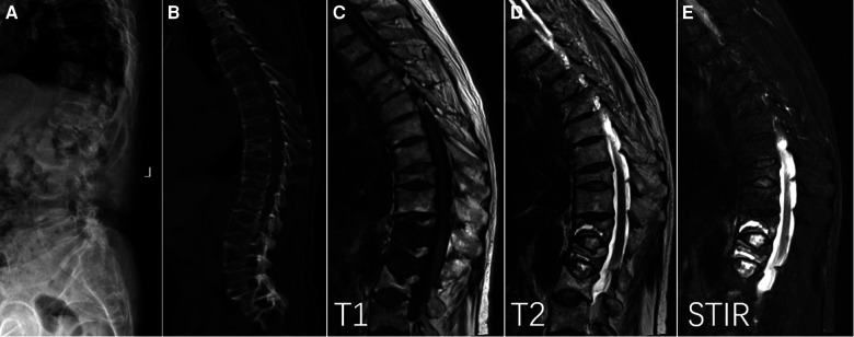
Figure 1.
Intravertebral vacuum phenomenon (IVP) with vertebral fractures and ankylosing spondylitis. Lateral x-ray (A) and sagittal CT (B) showed low-intensity cleft throughout T12 and L1, characterized by well-defined hypointensity on T1 (C), hyperintensity on T2 (D), and hyperintensity on short time inversion recovery (E).

