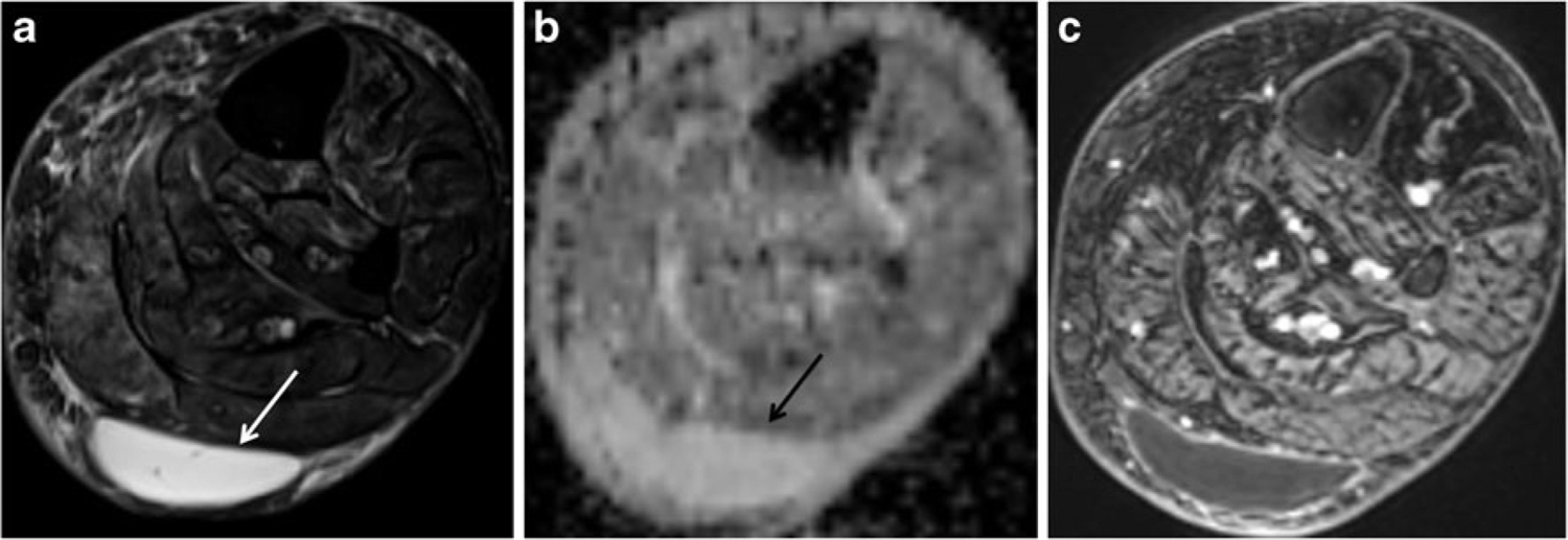
Fig. 1.

A 59-year-old woman with a posterior calf mass that resolved with follow-up. a Axial T2-weighted image with fat suppression (FSE, TR/TE 4,060/71 ms) shows a well-defined hyperintense mass (arrow) in the superficial soft tissues of the calf posterior to the gastrocnemius muscles. b Axial ADC map reveals the mass to have high ADC values (mean ADC value of 2.82). c Post-contrast image (VIBE, TR/TE 6.35/1.48) clearly shows the non-enhancing soft tissue mass (STM) as a cyst
