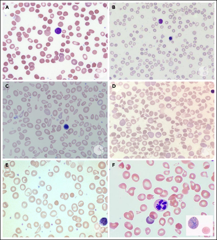
Figure 4.
PB smears. PB smears from (A) hereditary spherocytosis. Dense, spherical-shaped erythrocytes are seen. (B) β-Thalassemia major. Hypochromic, microcytic erythrocytes, anisocytosis, and a nucleated red blood cell are seen. (C) Sideroblastic anemia. Polychromasia, anisopoikilocytosis, and basophilic stippling are seen in a case of X-linked congenital sideroblastic anemia. (D) Thrombotic thrombocytopenic purpura. Anisopoikilocytosis and marked schistocytosis are seen on the smear of an infant with Upshaw-Schulman syndrome. (E) Iron deficiency. Significant anisocytosis, hypochromia, and microcytosis are seen. (F) Vitamin B12 deficiency. Macro-ovalocytes, microcytes, and hypersegmented neutrophils are seen. Erythrocyte basophilic stippling is shown in inset. These images were originally published in the ASH Image Bank. (A) Teresa Scordino, Hereditary spherocytosis, 2016, #00060308. (B) Girish Venkataraman, β-thalassemia major, 2018, #00062081; (C) Katherine Calvo, Congenital sideroblastic anemia peripheral blood, 2015; #00060064; (D) Helle Borgstrøm Hager and Mari Tjernsmo Andersen, Thrombotic thrombocytopenic purpura, #00061402; (E) Iron deficiency anemia moderate, 2015, #00060219. (F) Volodymyr Shponka and Maria Proytcheva, Megaloblastic anemia caused by severe B12 deficiency in a breastfed infant. 2017, #00061082. © The American Society of Hematology.

