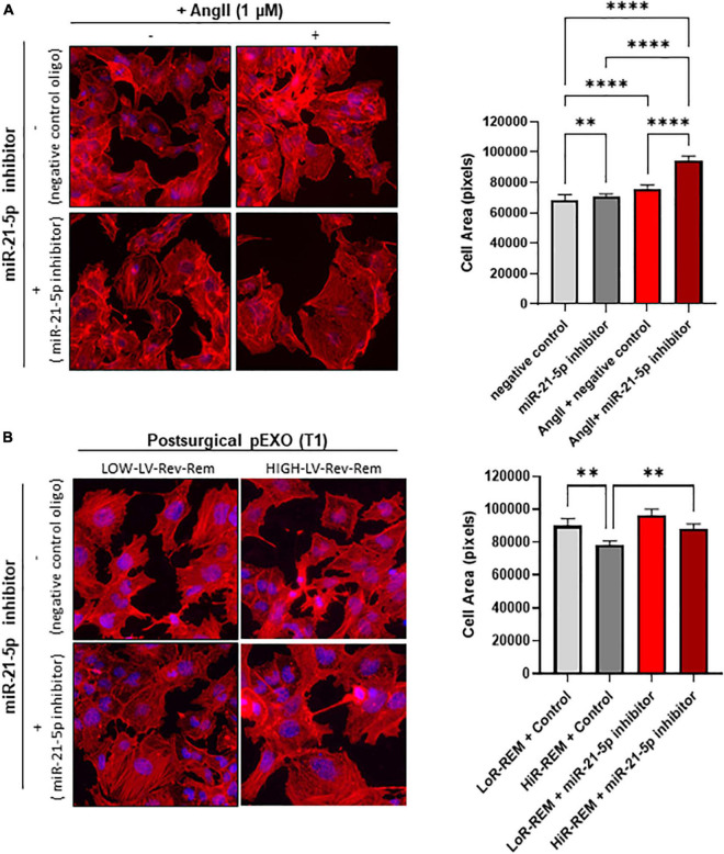
FIGURE 7.
Effect of miR-21-5p inhibition on cardiomyocytes size. The HL-1 cells were seeded at a concentration of 5,000 cells/cm2 and allowed to adhere for 24 h before treatment. (A) The HL-1 cells were treated with ± 1-μM angiotensin II (AngII, Sigma) in complete antibody-free Claycomb medium for 48 h. Moreover, 24 h after the beginning of the treatment, 30-nM miRNA-21-5p inhibitor/negative control was added to the cells using Lipofectamine® RNAiMAX Transfection Reagent and transfection was maintained for the following 24 h. (B) To stimulate hypertrophy, HL-1 cells were treated with 1-μM angiotensin II (AngII, Sigma) in complete antibody-free Claycomb medium for 48 h. The pEXOs isolated at T1 from LoR-REM or HiR-REM patients were loaded with miRNA-21-5p inhibitor/negative control through a heat shock-mediated protocol, as described in the experimental procedure, then collected by ultracentrifugation. Isolated pEXOs were added at a final concentration of 1 × 10⁹ particles/ml 24 h after the beginning of the AngII treatment, and treatment by pEXOs was maintained for the remaining 24 h in the presence of AngII. (A,B) At the end of each treatment, the cell monolayer was stained with Phalloidin–Atto 550 as described in the methods. The images were acquired with a fluorescence microscope (Leica; 20× magnification); the cell areas were manually measured with ImageJ software (https://imagej.nih.gov/ij/) and expressed in pixels. Statistical significance of the differences between treatments was evaluated by one-way ANOVA. **p < 0.01; ****p < 0.0001.

