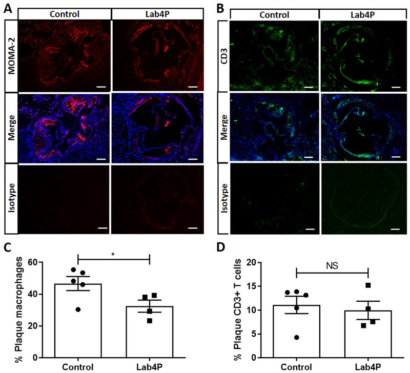
Figure 2.

Lab4 produces a significant decrease in plaque macrophage content in LDLr−/− mice fed HFD. Immunohistochemistry (IHC) of sections from the aortic root of mice fed HFD for 12 weeks (Control) or HFD supplemented with Lab4P (Lab4P) were stained using the MOMA‐2 antibody or CD3 antibody (both mounted with DAPI) or appropriate isotype control antibodies and images taken by fluorescent microscopy. Representative images are shown for control and Lab4P groups for MOMA‐2 staining (MOMA‐2), MOMA‐2 and DAPI staining (Merge) and IgG isotype control (Isotype) (A) or CD3 staining (CD3), CD3 and DAPI staining (Merge) and IgG isotype control (Isotype) (B) (scale bars 50 µm; red, MOMA‐2 AF‐488; green, CD3 AF‐594; blue, DAPI). The plaque content of macrophages (C) or CD3+ T cells (D) were determined using the Image J software. Data are mean ± SEM (n = 5 for Control and 4 for Lab4P). Statistical analysis was carried out using an unpaired Student's t test (*p ≤ 0.05).
