Fig. 3.
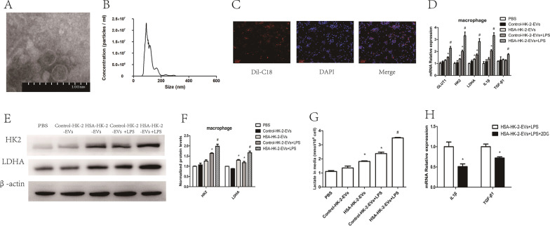
HSA-treated tubular epithelial cell-derived EVs promoted macrophage glycolysis in vitro. A EV morphology was analyzed using transmission electron microscopy. B EV size distributions were analyzed using nanoparticle tracking analysis. C EVs labeled with Dil-C18 were taken up by macrophages (original magnification ×100). Macrophages were co-cultured with EVs: D mRNA levels of GLUT1, HK2, LDHA, IL1β and TGF-β1 (n = 3); E, F protein levels of HK2 and LDHA (n = 3); G amount of lactate in the macrophage supernatant (n = 3); *p < 0.05 vs. the Control-HK-2-EVs group; #p < 0.05 vs. the Control-HK-2-EVs + LPS group. Macrophages treated with or without 2-DG were co-cultured with HSA-HK-2-EVs: H mRNA levels of IL1β and TGF-β1 (n = 3); *p < 0.05 vs. the HSA-HK-2-EVs + LPS group

