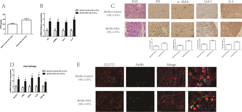
Fig. 4.
HSA-treated tubular epithelial cell-derived EVs promoted macrophage glycolysis in vivo. db/db mice were injected with Control-HK-2-EVs and HSA-HK-2-EVs via the caudal vein: A urinary albumin creatinine ratios (ACRs) of db/db + Control-HK-2-EVs (n = 5) and db/db + HSA-HK-2-EVs (n = 5) mice; B mRNA expression of FN, α-SMA, Col-I and IL-6 in the kidney cortexes of db/db + Control-HK-2-EVs (n = 5) and db/db + HSA-HK-2-EVs (n = 5) mice; C PAS staining and immunostaining of FN, α-SMA, Col-I and IL-6 (original magnification ×400) in the kidney cortexes of db/db + Control-HK-2-EVs (n = 5) and db/db + HSA-HK-2-EVs (n = 5) mice; D mRNA expression of GLUT1, HK2, LDHA, IL1β and TGF-β1 in kidney macrophages of db/db + Control-HK-2-EVs (n = 5) and db/db + HSA-HK-2-EVs (n = 5) mice; E double IHC staining of GLUT1 (red) and F4/80 (green) in the kidney cortex; *p < 0.05 vs. the db/db + Control-HK-2-EVs group

