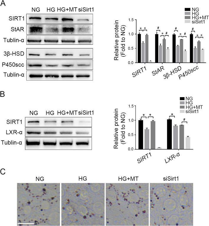
Fig. 4.
Sirt1 knockdown abolished melatonin protection of steroidogenesis against high glucose damages. A The protein levels of SIRT1, StAR, 3β-HSD and P450scc in TM3 cells were detected by Western blot and gray values were analyzed. B Western blot detection of SIRT1 and LXR-α expression in TM3 cells and gray value analysis of the protein bands. C Oil red staining was used to detect lipid droplets in TM3 cells. NG, normal glucose control group; HG, high glucose treatment group; HG + MT, high glucose and melatonin treatment group; siSirt1, HG + MT + siSirt1 group. A-B, Data are expressed as fold change relative to NG. *, P < 0.05; #, P < 0.01. Bar: C, 50 μm

