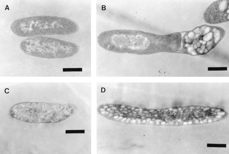
FIG. 5.
Electron micrographs demonstrating the sizes and localizations of PHB granules in recombinants of E. coli. The cells were cultivated at 37°C for 30 h in LB medium with sodium lactate, ampicillin, and kanamycin. (A) E. coli XL1-Blue (pBBRKmAB plus pTV119N); (B) E. coli XL1-Blue (pBBRKmAB plus pTVC); (C) E. coli XL1-Blue (pBBRKmAB plus pTVCP); (D) E. coli XL1-Blue (pBBRKmAB plus pTVCP4). Bars, 0.5 μm.

