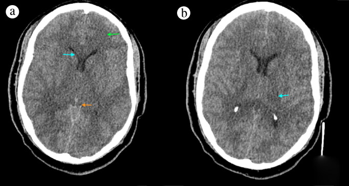
Figure 1.
Noncontrast CT (a) at the level of the basal cistern revealed extensive hypodensity in the white matter, basal ganglia, and thalami consistent with a global insult (green arrow), as well as narrow ventricles (blue arrow) and effacement of the basal cisterns (orange arrow). The (b) thalamus level showed mild hypodensity on the left thalamus.

