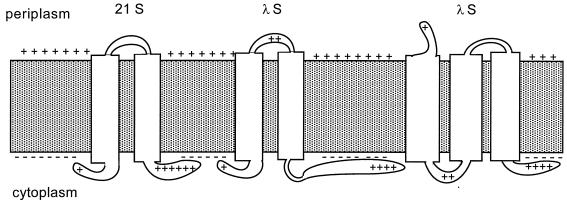
FIG. 6.
Possible membrane topologies of S. Depicted are putative membrane topologies for prototype class II (S from phage 21; left) and class I (S from phage λ; middle and right) holin proteins. The inner membrane is shown as shaded area with positively charged periplasmic and negatively charged cytoplasmic surfaces. Transmembrane helical domains are represented as white rectangles spanning the membrane. Basic residues in putative solvent-exposed domains are shown for both 21 and λ holins.

