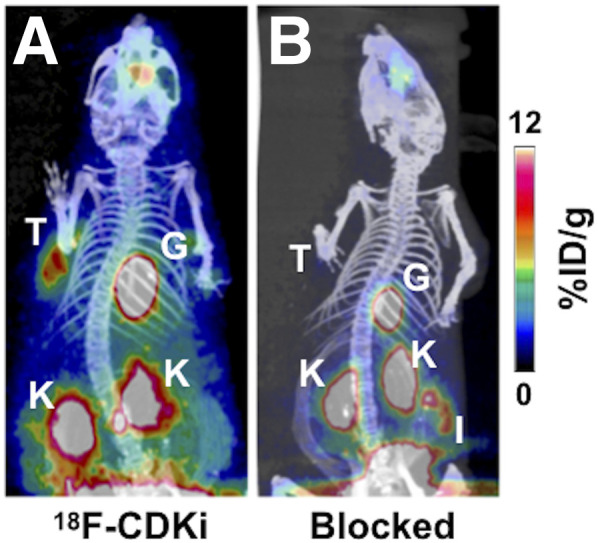
FIGURE 3.

In vivo small-animal PET/CT images of MCF-7–bearing mouse models. (A) Single-bolus injection of 18F-CDKi (7.4 MBq acquired at 2 h after injection). (B) Control MCF-7 mouse (preinjected with excess of palbociclib 30 min before 18F-CDKi, 7.4 MBq, with image acquired 2 h after injection). Active areas in scans are nasal epithelium and Harderian glands, tumor (T), gallbladder (G), kidneys (K), and intestines (I).
