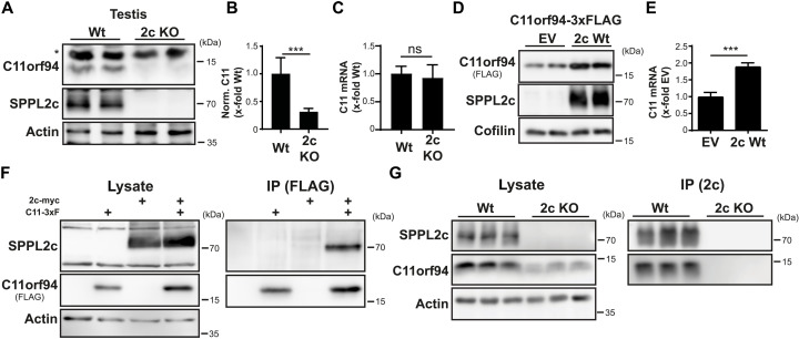
Fig. 2. C11orf94 physically interacts with SPPL2c.
(A) Protein levels of C11orf94 in the testis of either Wt or SPPL2c-deficient (2c KO) mice were determined by Western blotting. The asterisk highlights an unspecific band. (B) Quantification of (A). N = 2, n = 6, two-tailed unpaired Student’s t test. (C) Expression of the C11orf94 mRNA was analyzed by quantitative PCR using testis cDNA from Wt or Sppl2c−/− mice. N = 1, n = 7 (Wt)/6 (2c KO), two-tailed unpaired Student’s t test. (D) HEK cells were transiently transfected with C11orf94-3xFLAG alone or together with Sppl2c-myc. Protein levels were visualized by Western blotting. (E) Quantification of (D). N = 4, n = 8, two-tailed unpaired Student’s t test. (F) HEK cells were transfected with the indicated constructs. After lysis with 1% Triton X-100, pulldown of C11orf94 from the respective lysates was performed using anti-FLAG and protein G agarose beads. Lysates and bead eluates were subjected to Western blot analysis using the indicated antibodies. (G) Testes from wild-type or Sppl2c−/− mice were lysed in 0.5% CHAPSO. SPPL2c was precipitated from the lysates using a specific antibody and protein G agarose. Lysates and bead eluates were analyzed by Western blotting. ns, not significant. ***P ≤ 0.001.

