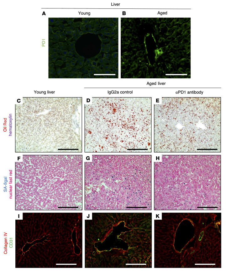
Figure 10. Anti–PD-1 antibody decreases liver aging in mice.
(A and B) Representative images of PD-1 immunofluorescence staining, which is higher in aged mouse liver (B) compared with young liver (A). (C–E) Oil Red staining (red) as a marker of fat deposition was barely detected in young livers but was increased in the livers of aged IgG2a-injected mice and decreased in aPD1ab-injected mice. (F–H) SA-β-gal staining (blue) used as a marker of senescence was increased in aged IgG2a-injected livers and decreased by aPD1ab treatment. (I–K) Double immunostaining for collagen IV (red) and the endothelial cell marker CD31 (green) shows increased collagen IV deposition in the blood vessels of the liver from aged IgG2a-injected mice compared with young mice, which was decreased by aPD1ab injection. Scale bars represent 50 μm (A and B) and 100 μm (C–K).

