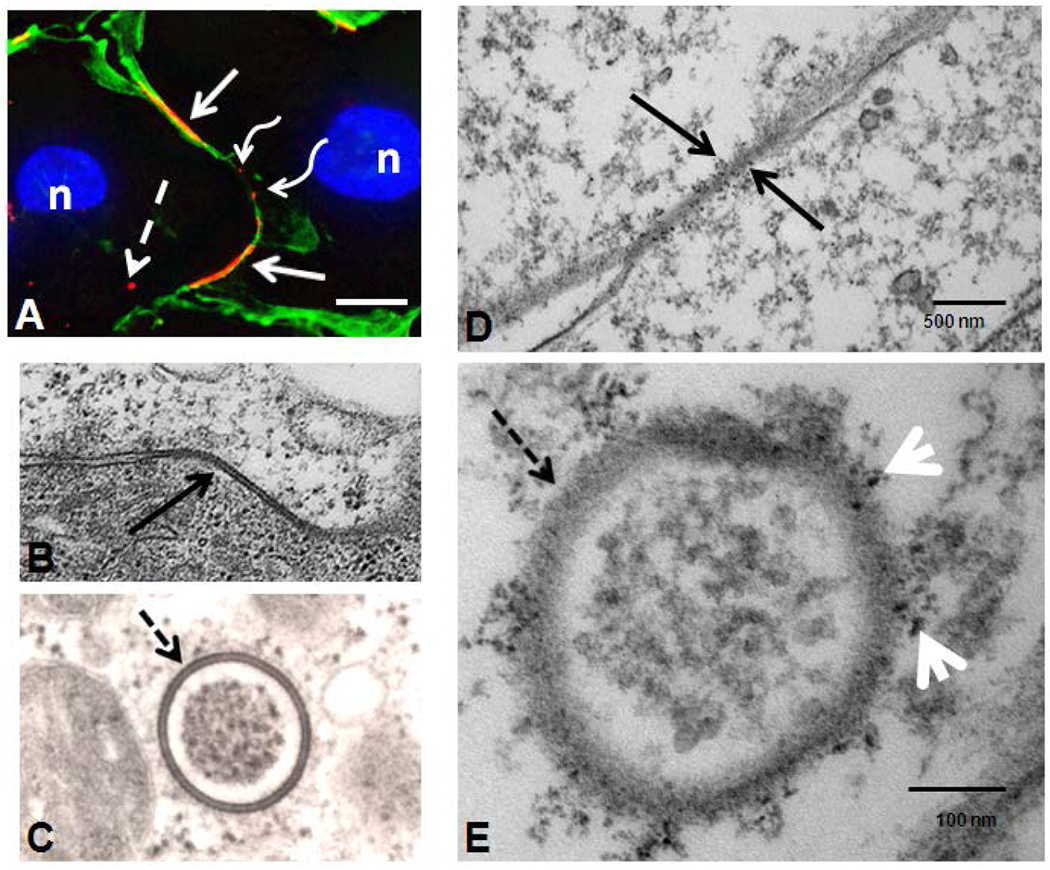
Figure 5.

Gap junction plaques (solid arrows) and annular gap junction (dashed arrows) seen in immunofluorescence (A), standard transmission electron microscopy (B,C) and quantum dot immuno-electron microscopy (D, E). Both large (solid arrow) and small gap junction plaques (waved arrows) can be seen on the cell surface (A). Cortical actin was stained with phalloidin Alexa-488 (green) to help define the cell borders and distinguish the cytoplasmic punctate staining (dashed arrow) from small surface gap junction plaques (waved arrow)(A). Note the typical pentalaminar membrane of the gap junction plaque (solid arrow in B) and annular gap junction vesicle (dashed arrow in C). The association of phosphorylated Cx43 with a gap junction plaque (solid arrow in D) and an annular gap junction vesicle (dashed arrow in E) has been demonstrated with Q-dot immuno-electron microscopy labeling. In E arrowheads are pointing to Cx43 Q-dots. Bars: 10μm A; 100nm B; and 100nm C; 500nm D; and 100nm E.
