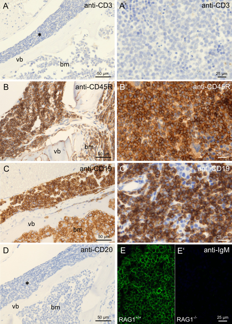
Fig. 3.
Immunohistochemical evaluation of lymphoid markers in cellular infiltrates in paralysis-affected RAG1-deficient mice. Immunohistochemical stainings of spine with cellular infiltrates in the spinal canal (asterisk) and the bone marrow cavity (bm) of vertebral bones (vb) and spleen of RAG1-deficient animals with hind limb paralysis. Anti-CD3 staining in spine A and spleen A′, anti-CD45R staining in spine B and spleen B′, anti-CD19 staining in spine C and spleen C′, anti-CD20 staining in spine D, anti-sIgM staining in wildtype animals E and paralysis-affected RAG1-deficient animals E′

