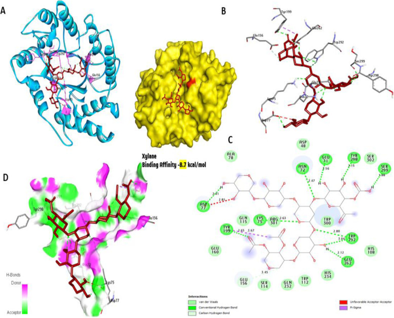
Fig. 24.
Molecular interactions of xylan with amino acids of the 3D model of xylanase with the best binding mode in the pocket of protein (with ligand as color sticks). A Complex interaction with sticks red color, B 3D interaction amino acid residues involved in the interaction (with ligand as color sticks), C 2D interaction binding interaction of ligands with an amino acid with hydrogen bond (green dash line), and D display receptor surface with H-bond donor and acceptor

