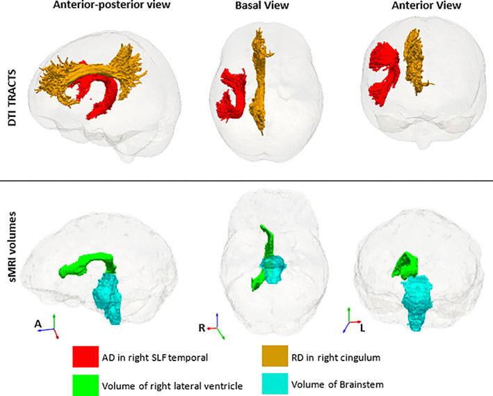
FIGURE 4|.

Four extra features that appear in the multimodal analysis and were not part of the single modality analyses of sMRI volumes and DTI. These are in addition to all except three (right choroid plexus and ventral diencephalon, and posterior corpus callosum) of the sMRI features in Figure 2 and the DTI tracts, except left corticospinal tract, in Figure 3. The DTI regions of interest are from the John Hopkins University (JHU) atlas (Mori et al., 2005) and the sMRI volumes from FreeSurfer (Fischl et al., 2002).
