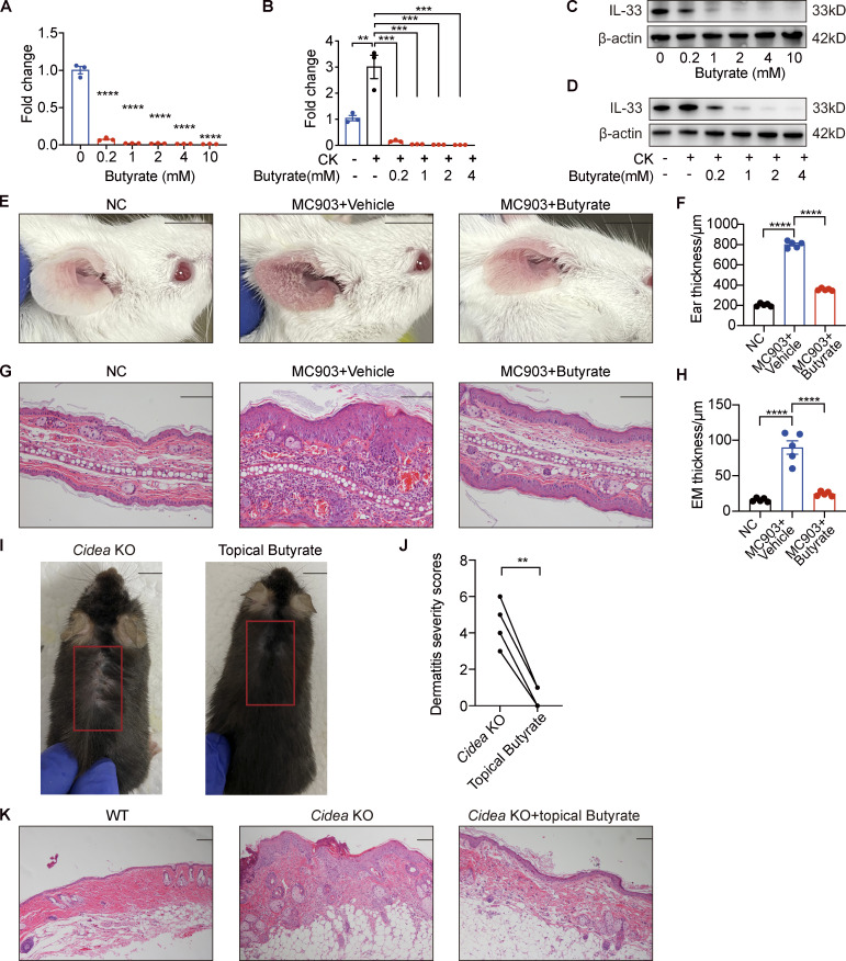
Figure S4.
Butyrate suppresses IL-33 expression in keratinocytes and inhibits skin inflammation of AD mouse model. (A–D) IL-33 mRNA and protein expression in keratinocytes treated with different concentrations of butyrate (0.2–10 mM) in standard culture medium or supplemented with IL-4 and IL-1α for 24 h (n = 3 per group). (E–H) MC903 or MC903 plus butyrate (1 mM)/vehicle was applied topically on the ears of BALB/c mice once daily for 9 d (n = 5 per group). (E) Representative gross appearance of the ears. (F) Ear thickness of the mice in each group. (G) H&E staining of ear sections. (H) Epidermal (EM) thickness of ear sections under high-power magnification. (I–K) Cidea KO mice were topically applied with butyrate (1 mM) on the lesional skin twice daily for 21 d (n = 4 per group). (I) Gross appearance of skin lesions in Cidea KO mice before and after topical butyrate treatment. The red box indicates the skin lesions treated with butyrate. (J) Severity scores of skin lesions in Cidea KO mice before and after butyrate treatment. (K) H&E staining of skin samples from WT mice, lesional skin of Cidea KO mice, and lesional skin of Cidea KO mice treated with butyrate. Scale bar = 1 cm (E and I); 100 μm (G and K). Data are representative of three independent experiments and are expressed as means ± SEM. Statistical significance was analyzed by one-way ANOVA followed by Dunnett’s test (A and B) and paired t test (J). *, P < 0.05; **, P < 0.01; ***, P < 0.001; ****, P < 0.0001. Source data are available for this figure: SourceData FS4.

