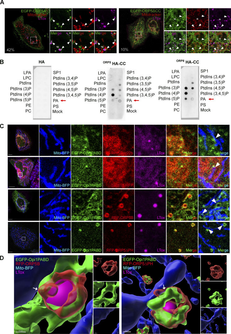
Figure 5.
ORP5 localizes to LDs and ER subdomains enriched in phosphatidic acid (PA). (A) Confocal images (single focal plane of HeLa cells expressing EGFP-tagged ORP5A or ORP5∆CC (green), treated with OA (300 μM) for 2 h. The mitochondria and the LDs were stained with Mitotracker (red) and LTox (purple), respectively. Arrowhead points ORP5-labeled MAM-LD associated with mitochondria and asterisks marks ORP5 localized to reticular ER. Scale bar, 5 µm. (B) PIP strip overlay assay: PIP strips were incubated with either ORP5-HA CC or ORP8-HA CC or the HA peptide as a negative control and analyzed using the anti-HA antibody. LPA, lysophosphatidic acid; LPC, lysophosphocholine; PtdIns, phosphatidylinositol; PtdIns(3)P; PtdIns(4)P; PtdIns(5)P; PtdIns(3,4)P2; PtdIns(3,5)P2; PtdIns(4,5)P2; PtdIns(3,4,5)P3; PA, phosphatidic acid; PS, phosphatidylserine; PE, phosphatidylethanolamine; PC, phosphatidylcholine; S1P, sphingosine 1-phosphate. (C) Confocal images (single focal plane) of HeLa cells co-expressing EGFP-Opi1PABD (green) with Mito-BFP (blue) and either RFP-Sec22b (red), or Sec61β-RFP, or EGFP-ORP5B (red), or EGFP-ORP5∆PH. The LDs were stained with LTox (purple). Arrowheads points enrichment of Opi1PABD at Mito–MAM–LD contact sites. Scale bar, 10 μm (entire cell), or 3 μm (zoom). (D) 3D reconstruction of cells shown in D using IMARIS. Arrows point to the MAMs where ORP5B and ORP5∆PH co-localize with Opi1PABD at Mito–MAM–LD contact sites. Scale bar, 0.5 µm.

