Figure 7.
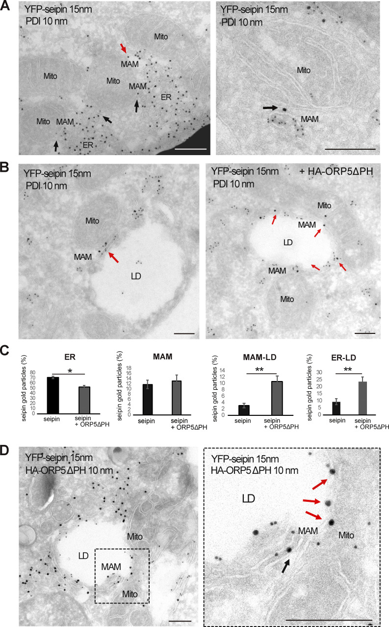
ORP5∆PH increases the localization of seipin to MAM–LD and ER–LD contacts. (A) Representative images of electron micrographs of ultrathin cryosections of HeLa cells transfected with YFP-seipin and immunogold stained with anti-GFP (15 nm gold) to detect seipin and anti-PDI (10 nm gold) to label the ER lumen. Seipin localizes at MAM–LD contacts (arrows). Mito, mitochondria; ER, endoplasmic reticulum; MAM, mitochondria-associated membranes; LD, lipid droplets. Scale bar, 250 nm. (B) Representative images of electron micrographs of ultrathin cryosections of HeLa cells co-transfected with YFP-seipin alone or together with HA-ORP5. Cells were immunogold stained with anti-GFP (15 nm gold) to detect seipin and anti-PDI (10 nm gold) to label the ER. (C) Quantification of the distribution of seipin immunogold particles (15 nm). Data are shown as % mean ± SEM of cell profiles with n = 32 (750 gold particles analyzed) in seipin individual expression, and n = 50 (940 gold particle) in seipin + ORP5∆PH co-overexpression. *P < 0.001, **P < 0.0001, unpaired two-tailed t test. (D) Electron micrographs of ultrathin cryosections of HeLa cells co-transfected with YFP-seipin and HA-ORP5∆PH and immunogold stained with anti-GFP (15 nm gold) to detect seipin and anti-HA (10 nm gold) to detect ORP5. The localization of seipin at MAM-LD contacts is increased when co-expressed with ORP5∆PH (arrows). Scale bar, 250 nm.

