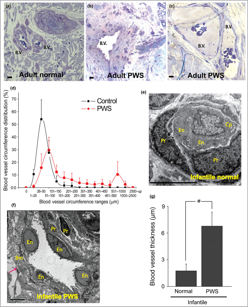
Fig 1.

Thick- and thin-walled blood vessels in adult and infant port-wine stain (PWS) lesions. (a) Normal blood vessels (b.v.) in adjacent normal skin from an adult with PWS. (b, c) Thick- and thin-walled PWS blood vessels from adults with PWS. (d) PWS blood vessel circumference distribution vs. normal dermal vasculatures (four patients with PWS and four normal adult participants). (e) Electron microscopy (EM) showed a normal capillary in adjacent normal skin from an infant with PWS. En, endothelial cell; Pr, pericyte; Cp, capillary. (f) EM showed an ectatic, thick-walled blood vessel with replication of the basement membranes in PWS from the same subject as in (e). Bm, basement membrane. The arrow indicates the blood vessel wall. (g) Infantile PWS blood vessels showed a significantly thicker blood vessel wall compared with normal dermal blood vessels from the same subjects (n = 4). Scale bar = 5 μm. *P < 0·05 vs. control.
