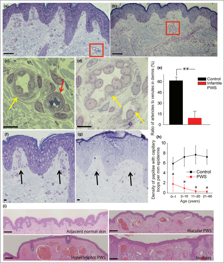
Fig 2.

Multiple developmental impairments of infant port-wine stain (PWS) vasculatures. (a) Semi-thin section showed normal adjacent skin from an infant with PWS. (b) Semi-thin section showed PWS lesional skin from the same subject as in (a). Scale bar = 20 lm. (c) A normal venule (yellow arrow) and arteriole (red arrow) from the red-boxed area in (a). (d) PWS pathoanatomical venule-like vasculatures (yellow arrows) from the red-boxed area in (b). Scale bar = 5 μm. (e) The ratio of arteriole to venule-like vasculatures in infantile PWS lesions was significantly reduced compared with normal adjacent skin from the same subjects (n = 4). (f) Normal formation of capillary loop (black arrows) in adjacent normal skin from an infant with PWS. (g) Defects in capillary loop formation along with normal development of epidermal rete ridges in PWS from the same subject as in (d). Scale bar = 5 μm. (h) Quantitative analysis of the density of papillae containing capillary loops per mm epidermis in patients with PWS vs. normal subjects among groups of different ages. (i) Reduction of capillary loops and rete ridges in PWS flat reddish macular, protuberant hypertrophic areas and nodules from the same subject. Scale bar = 100 μm. **P < 0·01 and *P < 0·05 vs. the control groups in (e) and (h).
