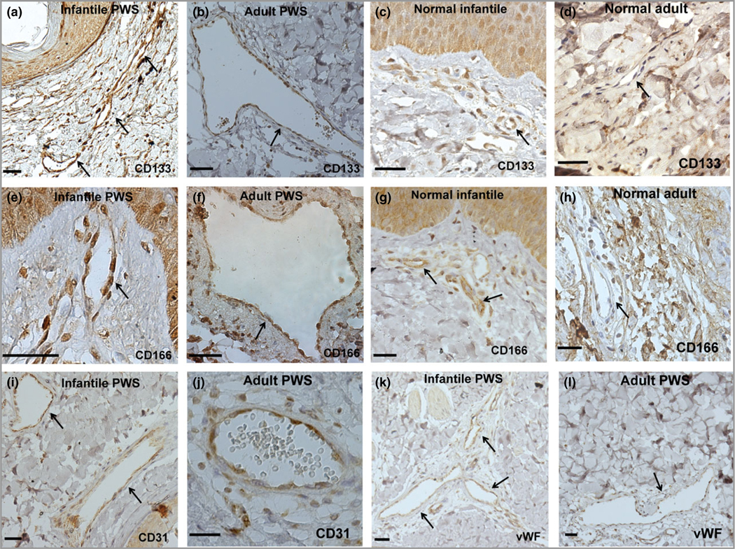
Fig 3.

Port-wine stain (PWS) endothelial cells (ECs) presented stemness phenotypes of CD133+/CD166+ in non-nodular lesions. (a–h) Expression of CD133 and CD166 in infant and adult PWS and normal subjects. (i–l) PWS ECs expressed EC markers CD31 and von Willebrand factor (vWF). Positive stain is diaminobenzidine (DAB) (brown). Scale bar = 50 μm. Arrows indicate blood vessels.
