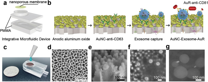
Fig. 10.
Integrative microfluidic device for exosome isolation and detection. a Design of the integrative microfluidic device. b Schematic illustration of in-situ detection of exosome. c A photographic image of the integrative microfluidic device. d SEM image of nanoporous gold (Au) nanoparticles deposited on AAO membrane with a thickness of 50 nm. e, f The side view of Au coating. g SEM image of the formed complex nanoporous gold nanocluster (AuNC)-Exosome-AuR. Reproduced with permission [60]. Copyright 2020, Elsevier

