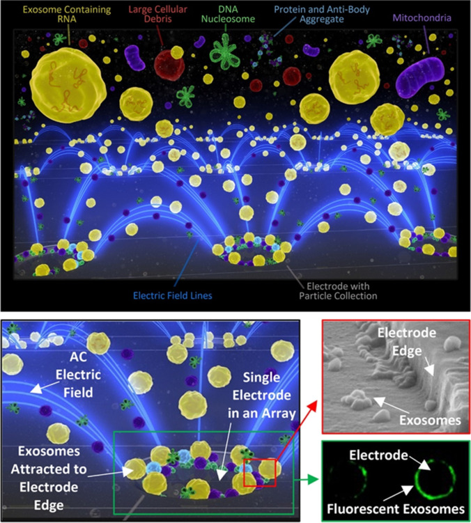
Fig. 11.
Schematic representation of exosome isolation on the ACE device (chip) microelectrodes. Electric field lines (blue) run between individual microelectrodes on the microarray and converge onto the edges of the microelectrodes, thereby forming the DEP high-field regions. The exosomes collect in the high-field regions while cells or larger particles in the sample are concentrated into the DEP low-field areas between microelectrodes, and lower molecular weight biomolecules are unaffected by DEP electric fields. A fluid wash removes any cells and the other plasma materials while the nanosize biomarkers (exosomes, etc.) remain concentrated in the DEP high-field regions. Reproduced with permission [68]. Copyright 2017, American Chemical Society

