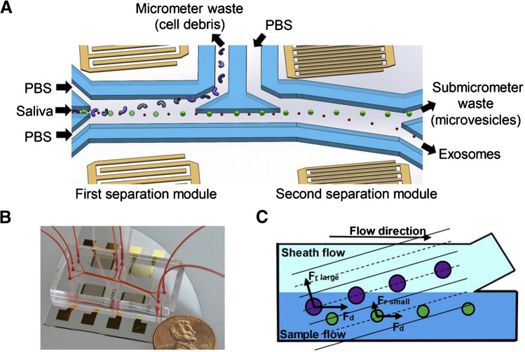
Fig. 13.
Schematic and mechanism of the acoustofluidic device for exosome separation. A, B Schematic and optical image of the acoustofluidic device (penny shown for size comparison). C There is a size-based separation in each module. Given the acoustic radiation force (Fr) and a drag force induced by fluid (Fd), larger particles are separated into the sheath flow while small particles remain in the primary sample flow. Reproduced with permission [75]. Copyright 2020, Elsevier

