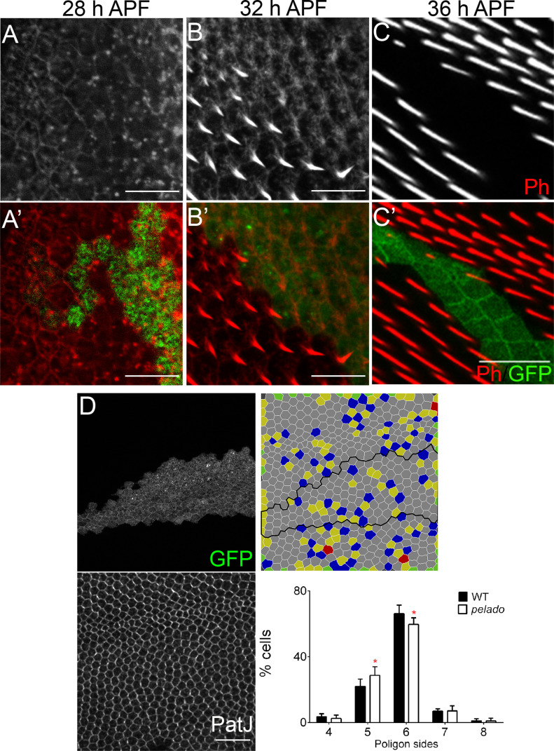
Figure 2. Pldo is required for actin hair formation and cell shape maturation.
(A, B, C) Temporal characterization of the mutant pldo phenotype during cellular hair formation. Confocal micrographs showing pldo MARCM clones (marked by GFP, green) during actin hair formation (labeled via rhodamine phalloidin, Ph). (A, B, C) Monochrome images showing actin alone (Ph) and (A′, B′, C′) displaying clonal marker (green) and actin (red). Note the absence of hair elongation from 30 to 32 h APF onward, in mutant GFP marked cells. (A, A′) Note accumulation of actin appears normal in control and mutant cells at 28 h APF. All the images are oriented with distal in the lower right corner. Scale bars correspond to 50 μm. (D) Evaluation of cell shape in pldo-mutant cells, at 28–32 h APF, as compared with neighboring wild-type cells. Note the significant reduction in the number of hexagonal cells in pldo-mutant clones (marked by GFP; cell outline displayed via PatJ staining, a junctional marker). Six different individuals were analyzed, with around 100 cells in each case. P = 0.0366 via t test. Scales bar correspond to 100 μm.

