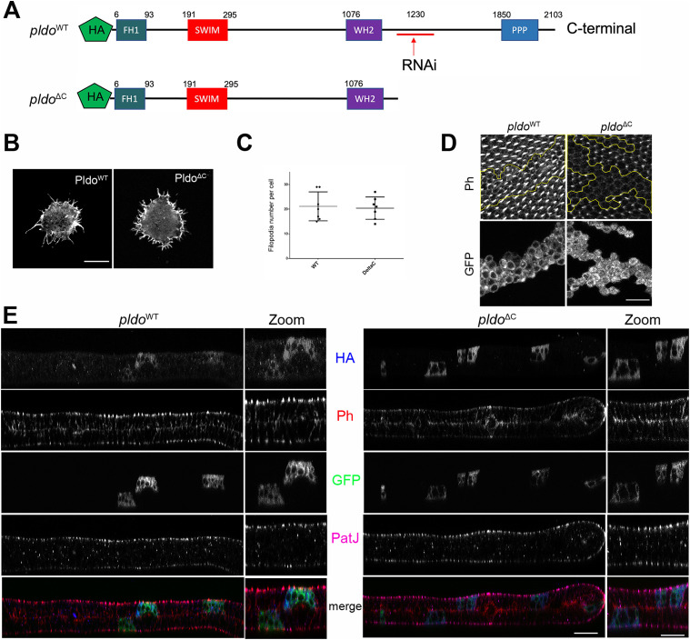
Figure 5. N-terminal region of Pldo is sufficient to induce filopodia formation.
(A) Schematic of Pldo highlighting the deletion isoform with C-terminal deletion, PldoΔC (note that the RNAi sequences are absent in the PldoΔC construct). Both constructs have an HA tag at the N-terminus. (B, C) PldoΔC was sufficient to induce formation of filopodia in hemocytes, to the same extent as full-length PldoWT. (B, C) Examples shown in (B) and quantification in (C), showing the number of filopodia per cell. Scale bar represents 10 μm. At least 10 cells were analyzed in each condition in three independent experiments. Statistical analysis with t test, P = 0.8028, nonsignificant. (D) Expression of PldoΔC in MARCM pldo-mutant cells (right panels) in pupal wings was not able to rescue hair formation, compared to pldoWT expression (left panels). Distal is right. Scale bar represents 25 μm. (E) Subcellular localization (z-plane) analyses of HA-tagged PldoWT (left panels) and PldoΔC (right panels), displaying rhodamin phalloidin (Ph, red) staining actin filaments, clonal marker (GFP, green, via MARCM, mutant cells), PatJ (magenta) as a junctional (apical) cell marker, and HA-Pldo (blue) showing Pldo localization. Note that there is no detectable difference in the localization of PldoWT and PldoΔC (HA staining, blue) in pupal wing cells. Right panels (zoom) show higher magnification. Scale bar represents 25 μm.

