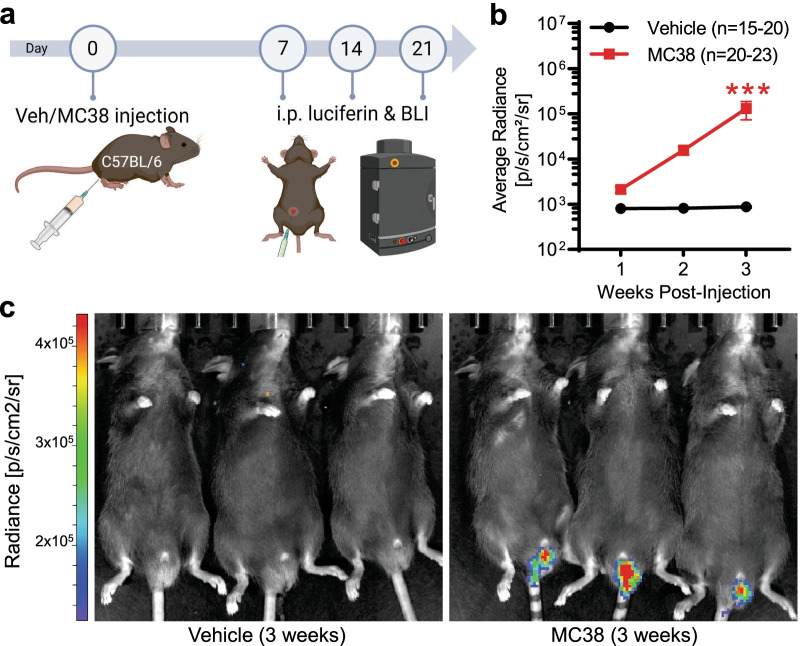
Fig. 1.
In vivo bioluminescence imaging of colon cancer development after local MC38 cell injection. a Schematic depicting of experimental protocol for IVIS bioluminescence measurements (animals were measured 7, 14, and 21 days after vehicle/MC38 cell injection). b Changes in average bioluminescence radiance values. The consistent localized increase in the measured bioluminescence signal reflects consistent tumor growth after transanal MC38 cell injection. ***: p < 0.001 vs MC38 week 1, mixed-effects model with Šídák's multiple comparisons test. c Representative image of three control (vehicle injected) and colon cancer bearing (MC38-injected) mice, 3 weeks after inoculation

