Fig. 8.
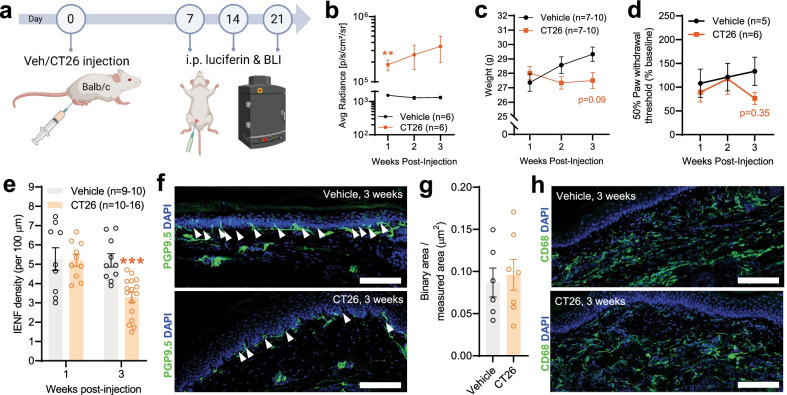
Tumor development, behavioral-, and IENF density changes in the CT26 colon cancer model. The data from animals with local injection of CT26 cells reveal very similar changes and patterns to the MC38 model. a Schematic representation of the experimental protocol for the IVIS in vivo bioluminescence measurements (animals were measured 7, 14, and 21 days after vehicle/CT26 cell injection). The consistent robust increase in the bioluminescence signal is depicted in b and indicates persistent colon cancer development (**: p < 0.01 vs vehicle-treated, two-way ANOVA with Šídák's multiple comparisons test). c No significant change in the development (animal weight, g) of mice with or without CT26 initiated colon cancer (mixed-effects analysis with Šídák's multiple comparisons test). d Von Frey testing of hind paws revealed no significant alteration in mechanical sensitivity. Datapoints represent the average of percentage changes, compared to the measured individual baseline values (measured at week 0, before inoculation. Two-way ANOVA with Šídák's multiple comparisons test). e–h Immunohistochemical analysis of hind paw samples: changes in IENF (PGP9.5) and macrophage (CD68) signals. e Changes in IENF density with time in hind paws of control vs CT26-injected mice. The IENF density in the hind paws of CT26-injected mice was significantly decreased compared to control (vehicle inj.), 3 weeks after tumor inoculation (***: p < 0.001, two-way ANOVA with Šídák's multiple comparisons test). f Representative images show the decrease in IENF density compared to control, 3 weeks after tumor inoculation. White arrows indicate intraepidermal nerve fibers. Scale bar: 100 μm. g No significant changes in CD68 signal intensity at the 3rd week in the hind paws of control vs CT26-injected mice (p = 0.73, two-tailed unpaired t test). h Representative images of hind paw samples show no change in the CD68 signal intensity of the epidermis (3 weeks after cell injection). Scale bar: 100 μm

