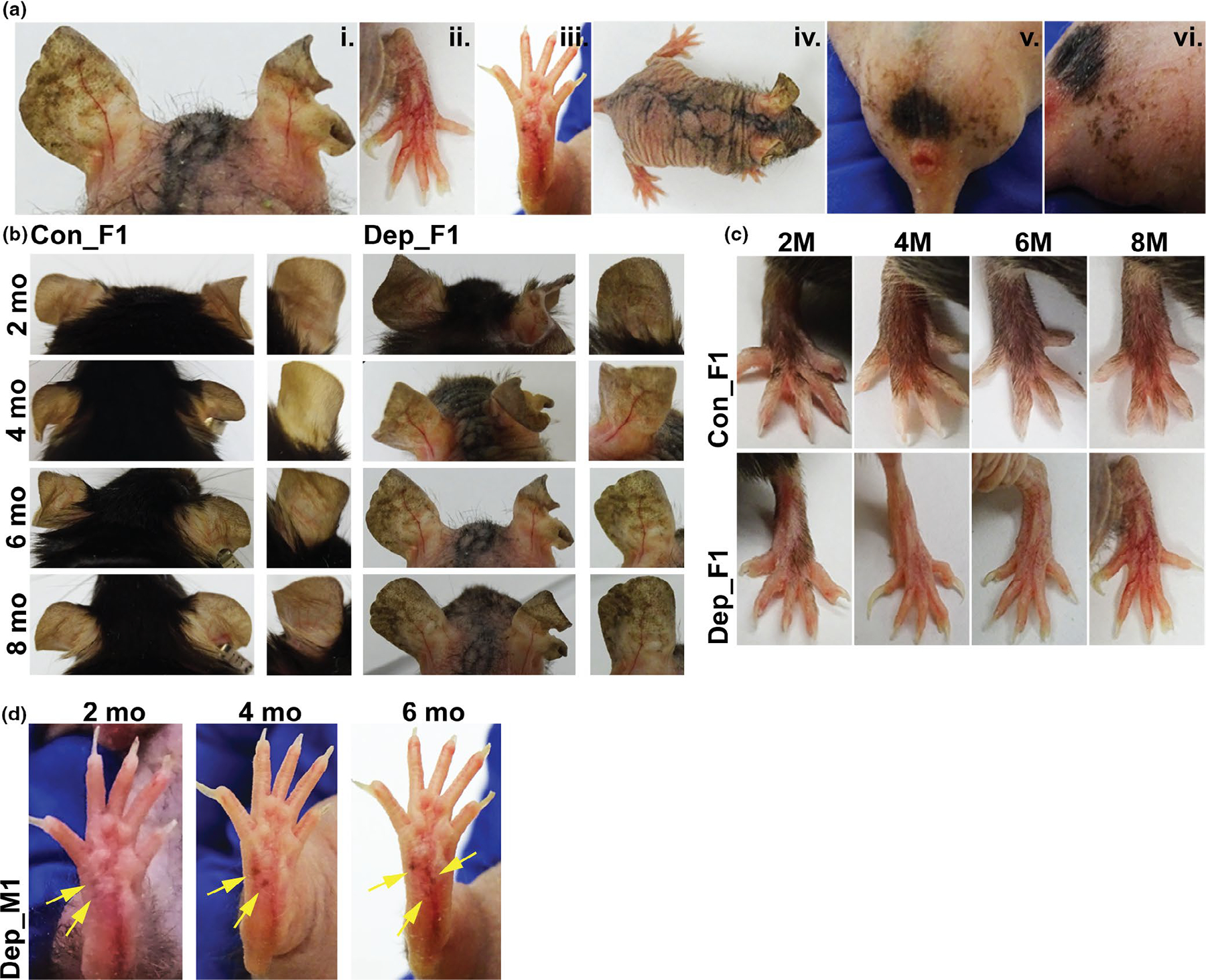
FIGURE 1.

Cutaneous pigmentary changes observed in mtDNA-depleter mice. (a) The spectrum of pigmentary phenotypes observed in the mtDNA-depleter mouse model. Representative images taken of mtDNA-depleter mice after induction with doxycycline diet showing patchy hyperpigmentation on the ear at 8 months (i), reticular hyperpigmentation on the dorsal surface of the paws at 8 months (ii), punctate brown pigmentation on the volar aspect of footpads at 6 months (iii), reticular hyperpigmentation on the dorsal skin surface of a female mouse at 2.5 months after healing of erythematous scaly rash (iv), and reticular hyperpigmentation on the genital and scrotal area of a male mouse that started after 6 months of doxycycline induction (v,vi). (b-c) A time course of changes in ear (b and paw (c) pigmentation in control (Con) and mtDNA-depleter (Dep) female mice. Representative images taken of mice over 2, 4, 6 and 8 months showing no or minimal changes in the ear pigmentation pattern in the control mice while the ears in mtDNA-depleter mice start to show diffuse hyperpigmentation as early as 2 months of doxycycline induction that turns into darker, patchy pigmentation over time. Control mice exhibit no change on the dorsal surface of the paws where they retain normal body hair, whereas the mtDNA-depleter mice experience hair loss followed by increased patchy and reticular hyperpigmentation over time. (d) Images of a male mtDNA-depleter mouse with ectopic pigmentation (yellow arrows) that appears at 2 months of doxycycline induction on the volar aspect of the paw that continues to erupt and darken over time
