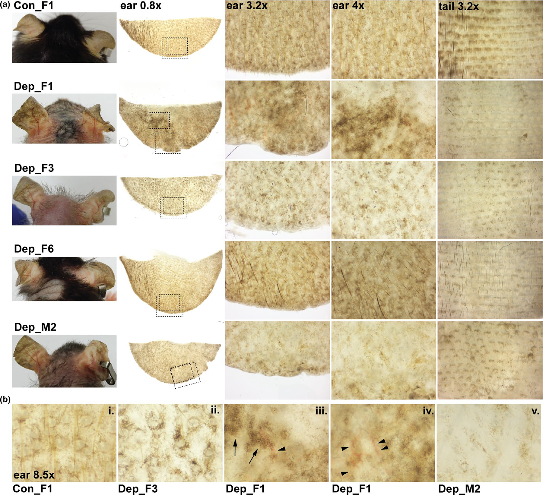
FIGURE 2.

Changes in ear and tail pigmentation in mtDNA-depleter mice. (a) Representative images of mice and whole-mount images of ears and tails harvested from control (Con) and mtDNA-depleter female (F) and male (M) mice after 8 months on doxycycline diet. Ears were imaged on their posterior side. Tails were images with the epidermis facing upward. The dashed boxes in the whole-mount ear 0.8× images indicate the magnified regions shown in the ear 3.2× and ear 4x higher magnification images. (b) High magnification images (8.5×) highlighting the spectrum of pigmentary changes observed when comparing control (i) with mtDNA-depleter mice (ii-v). Control mice display the normal pseudonetwork pattern of pigmentation including symmetrical pigmentation around adnexal openings, such as sebaceous glands and hair follicles (i). MtDNA-depleter mice exhibit accentuation of perifollicular, interfollicular pigmentation (ii); coalesced hyperpigmented patches with mottled areas of hyper and hypo pigmentation and dark brown dots (arrows, iii); linear pattern of blood vessels (arrowheads, iii-iv); and some areas with significant loss of pigmentation (v)
