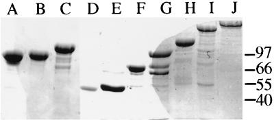
FIG. 2.
Denaturing gel electrophoresis of proteins expressed by the different plasmids. The proteins were stained with Coomassie brilliant blue. The structures of the epimerases were as follows: lane A, R3A2 (pHE51); lane B, A2R4 (pHE56); lane C, R3A2R4 (pHE27); lane D, A2 (pHE58); lane E, A1 (pHE42); lane F, A1R1 (pHE29); lane G, A1R1R2 (pHE35); lane H, A1R1R2R3 (pHE37); lane I, A1R1R2R3A2 (pHE57); lane J, A1R1R2R3A2R4 (pHH1). Six micrograms of protein was loaded in each lane. The proteins were partially purified by ion-exchange chromatography, except for the epimerase in lane B, which was further purified by gel filtration (11). The numbers indicate molecular masses (in kilodaltons) of a molecular mass standard.

