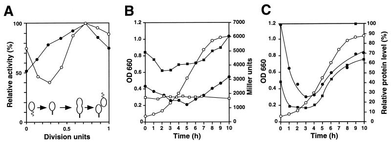
FIG. 7.
Cell cycle- and growth phase-dependent expression of clpP and clpX. (A) The relative activities of PX1 (○) and PX3 (●) were determined during the cell cycle by pulse labeling of synchronized cultures of strain NA1000 containing plasmids pAS6 (PX1) and pAS64 (PX3), respectively, with [35S]methionine for 5 min at different intervals and determining β-galactosidase synthesis during the pulse time by immunoprecipitation (see Materials and Methods). The promoter activity is shown relative to maximal activity. Progression of the cell cycle is indicated schematically at the bottom. (B and C) Growth phase-dependent expression (B) and cellular levels (C) of ClpP and ClpX. (B) Stationary-phase overnight cultures were diluted in fresh PYE complex medium (NA1000, NA1000/pAS2, and NA1000/pAS24) or PYE complex medium plus 0.2% xylose (NA1000/pCS225), and growth was monitored by determining the OD660 (○). At different intervals, samples were removed from the cultures, and β-galactosidase activity (Miller units) was determined as described in Materials and Methods for the following strains: NA1000/pAS2 (PP1) (●), NA1000/pAS24 (PX1, PX2, and PX3) (■), and NA1000/pCS225 (xylX promoter) (□). (C) Relative cellular levels of ClpP (●) and ClpX (■) in wild-type strain NA1000 were determined as a function of the growth phase by immunoblot analysis. ○, OD660.

