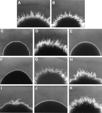
FIG. 1.
Colony morphology of various Y. lipolytica strains. (A and B) Wild-type strains E122 and 22301-3; (C) original mutant strain mhy1-1; (D) strain mhy1K09 transformed with plasmid pMHY1; (E and F) MHY1 disruptant strains mhy1K09 and mhy1K09-B4; (G) MHY1/MHY1 diploid strain E122//22301-3; (H and I) MHY1/mhy1 diploid strains 22301-3/mhy1K09 and E122//mhy1K09-B4; (J) mhy1/mhy1 diploid strain mhy1K09//mhy1K09-B4; (K) mhy1/mhy1 diploid strain mhy1K09//mhy1K09-B4 carrying plasmid pMHY1. The colonies were photographed at ×100 magnification after 3 days of incubation at 28°C on YNA agar plates.

