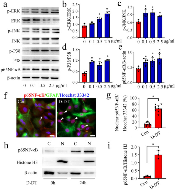
Fig. 5.

Effects of D-DT on the activation of intracellular MAPKs/NF-κB signaling in the astrocytes. a Western blot analysis of phosphorylation of ERK, P38, JNK kinase and p65NF-κB protein after stimulation of astrocytes with 0-2.5 µg/ml recombinant D-DT protein for 24 h. b–e Quantification data as shown in a. f Immunofluorescence showed the distribution of p65NF-κB in the cytoplasm and nucleus of astrocytes following treatment with 1 µg/ml recombinant D-DT for 24 h. Arrowheads indicate p65NF-κB-positive nucleus. g Quantification of p65NF-κB-positive nucleus co-stained with Hoechst 33,342 as shown in f. h Western blot analysis of p65NF-κB protein levels in the cytoplasm and nucleus following astrocytes treatment with 1 µg/ml recombinant D-DT for 24 h. i Quantification data as shown in h. Quantities were normalized to endogenous β-actin (cytoplasm) or Histone H3 (nucleus). Scale bar, 20 μm in f. Experiments were performed in triplicates. Error bars represent the standard deviation (*P < 0.05)
