
Fig. 3.
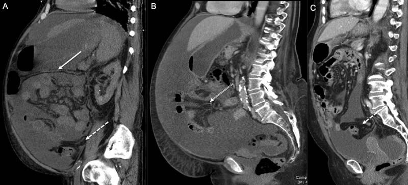
Panel of photographs generated from computerized axial tomographic imaging of the abdomen and pelvis ( A – C ) with intravenous contrast in the portal venous phase. The sagittal aspect of the abdomen is demonstrated showing a large volume of intraperitoneal free fluid. This fluid delineates the transverse mesocolon (solid arrow), small bowel region of the mesentery (double line arrow) and mesosigmoid (dashed arrow).