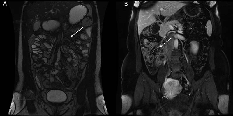
Fig. 4.

( A ) Photograph of T2 weighted coronal image of the of the abdomen generated from magnetic resonance imaging (MRI). The photograph demonstrates how MRI can be used to depict the anatomy of the small bowel region of the mesentery in normality (solid arrow). ( B ) Photograph of T1 fat-saturated coronal MRI with contrast demonstrating malrotation; the small bowel and adjoining mesentery are located on the right side of the abdomen (dashed arrow).
