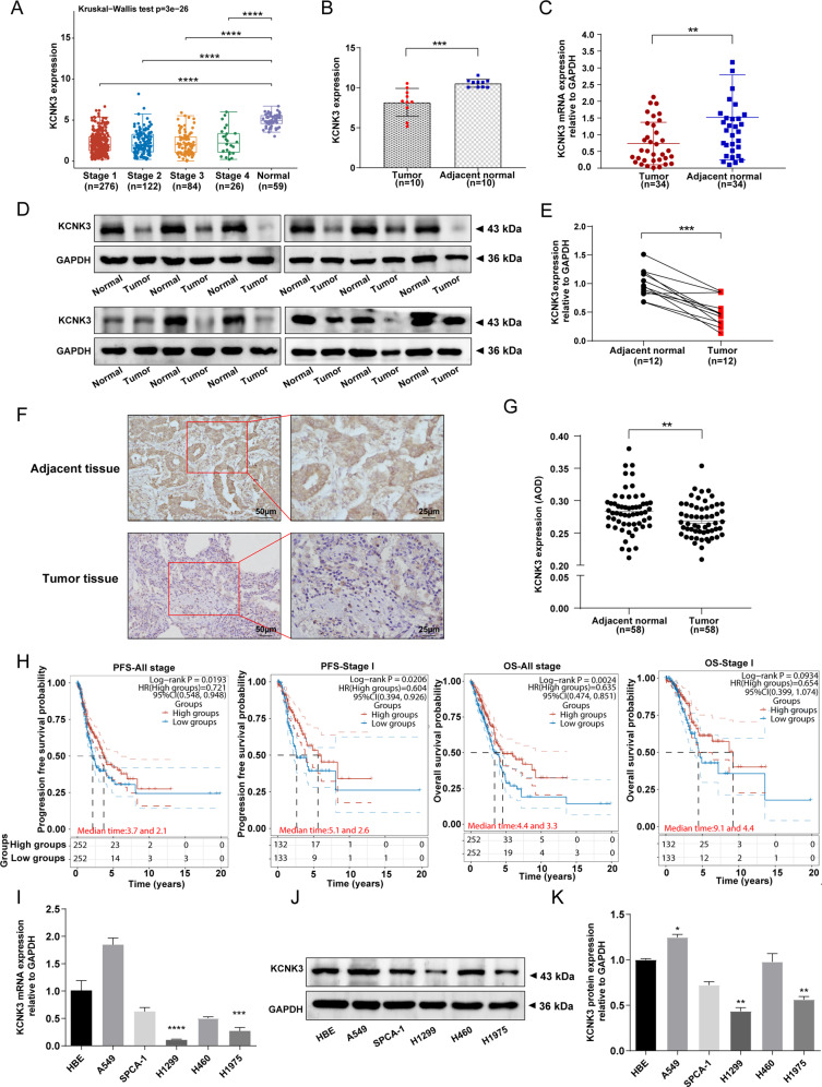
Fig. 1. Low KCNK3 expression in LUAD tissues was correlated with poor prognosis.
A The KCNK3 mRNA levels in LUAD tissues compared with the normal lung tissues from TCGA database. B, C The mRNA expression of KCNK3 in stage I LUAD patients was determined by RNA sequencing (n = 10) and qRT-PCR (n = 34). D, E Western blot analysis of KCNK3 protein expression in stage I LUAD tissues and corresponding adjacent tissues, with results normalized relative to the expression of GAPDH (n = 12). F, G KCNK3 protein expression in stage I LUAD tissues and adjacent tissues were detected by IHC and quantitative analysis by using average optical density (AOD, AOD = IOD/area, n = 58, scale bar 50 or 25 µm). H The prognostic values of KCNK3 in LUAD patients from TCGA by using a clinical bioinformatics database (www.aclbi.com). I–K KCNK3 mRNA and protein expression levels of LUAD cell lines were assessed by qRT-PCR and western blot, respectively. (OS, overall survival; PFS, progression-free survival. *P < 0.05, **P < 0.01, ***P < 0.001, ****P < 0.0001).

